Typical magnetic resonance imaging scan showing the coracohumeral
Por um escritor misterioso
Last updated 23 dezembro 2024


Typical magnetic resonance imaging scan showing the coracohumeral

Presentation1, radiological imaging of adhesive capsulitis(frozen shoulder).

The Rotator Interval: A Review of Anatomy, Function, and Normal and Abnormal MRI Appearance
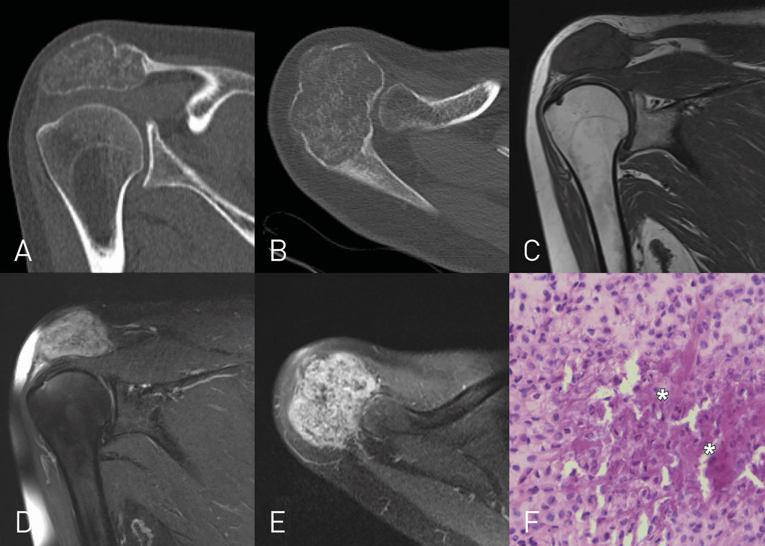
A 38-Year-Old Man with Long-Term Shoulder Pain - JBJS Image Quiz

Glenohumeral Instability
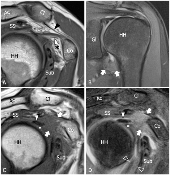
Diagnostics, Free Full-Text
:watermark(/images/watermark_5000_10percent.png,0,0,0):watermark(/images/logo_url.png,-10,-10,0):format(jpeg)/images/overview_image/903/qMKVkPzQ2XBi3WLkpgHGWQ_mri-pd-axial-glenoid-cavity-level_english.jpg)
Normal shoulder MRI: How to read a shoulder MRI
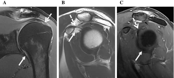
MR imaging detection of adhesive capsulitis of the shoulder: impact of intravenous contrast administration and reader's experience on diagnostic performance

Shoulder Anatomy and Normal Variants - Journal of the Belgian Society of Radiology
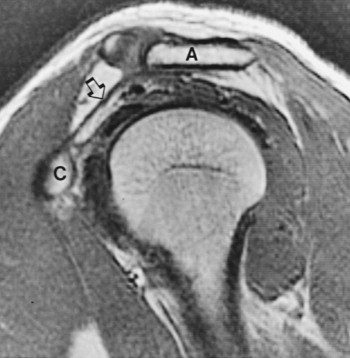
Shoulder Radiology Key
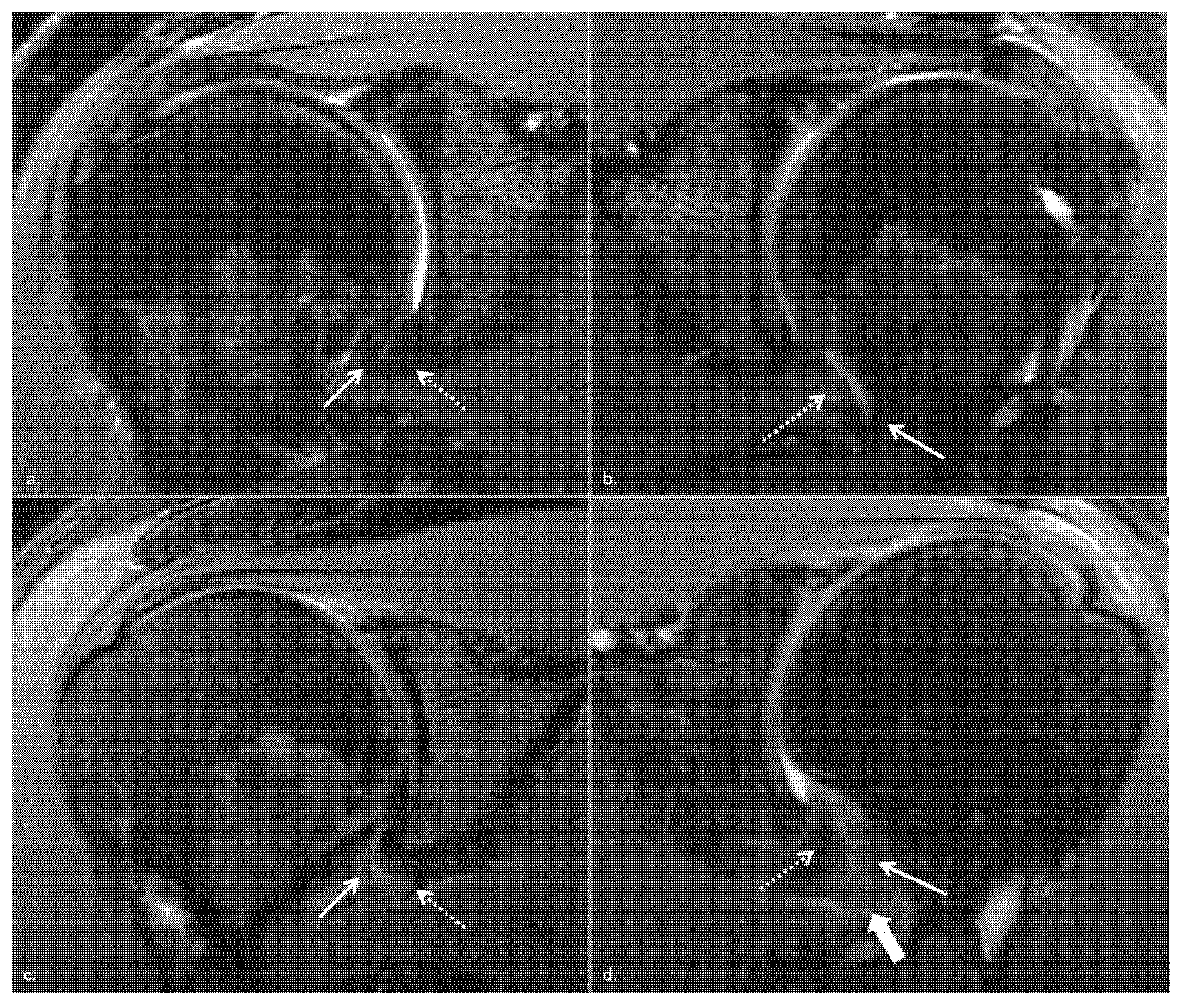
JCM, Free Full-Text

Alteration of coracoacromial ligament thickness at the acromial undersurface in patients with rotator cuff tears - ScienceDirect
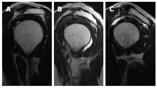
Rotator cuff disorders: How to write a surgically relevant magnetic resonance imaging report?
Recomendado para você
-
 SCP 007-J, Object Class: Euclid (Awaiting Keter Classification)23 dezembro 2024
 -
 900+ SCP Foundation ideas in 202323 dezembro 2024
 -
 LilyFlower's Workbench - SCP Foundation23 dezembro 2024
 -
 SCP SONGS VOL. 123 dezembro 2024
 -
 049 and his lil brother 049-J how cute23 dezembro 2024
 -
 𝟖𝐓𝐇 𝐏𝐑𝐎𝐉𝐄𝐂𝐓 🟧 on X: We are @TheCryptomasks 🎭 Sold for 0.3 E. / X23 dezembro 2024
 -
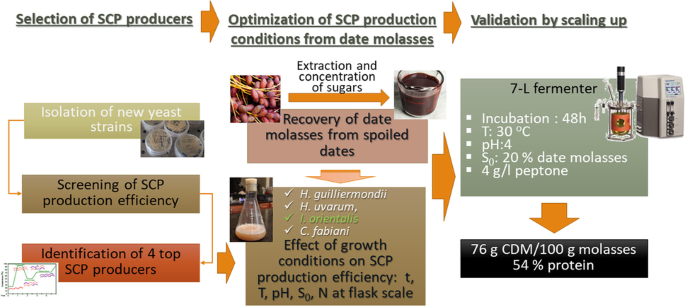 Valorizing food wastes: assessment of novel yeast strains for enhanced production of single-cell protein from wasted date molasses23 dezembro 2024
 -
 SCP-S4S – SCP-173-J Song (The Sculpture) Lyrics23 dezembro 2024
 -
Empty_I's (@empty_i.s) • Instagram photos and videos23 dezembro 2024
-
 Mitochondrial defects caused by PARL deficiency lead to arrested spermatogenesis and ferroptosis23 dezembro 2024
você pode gostar
-
 Ore No Kanojo To Osananajimi Ga Shuraba Sugiru Wallpapers - Wallpaper Cave23 dezembro 2024
 -
Is the original Ridley Scott-directed science-fiction horror film23 dezembro 2024
-
/cdn.vox-cdn.com/uploads/chorus_image/image/72108951/Screen_Shot_2022_07_23_at_9.44.08_AM.0.png) John Wick 4's post-credits scene proves the action never really23 dezembro 2024
John Wick 4's post-credits scene proves the action never really23 dezembro 2024 -
 Character Art: Super Sonic23 dezembro 2024
 -
 JOJO's Bizarre Adventure Part.6 Stone Ocean Deco Sticker Jotaro Kujo Japan rare23 dezembro 2024
 -
 Kingdom Clash Wiki23 dezembro 2024
 -
 FromSoftware Reportedly Working on Elden Ring 2 to Counter and23 dezembro 2024
 -
 Download Follow the adventure of Kazuma and his mischievous gang in the world of Konosuba.23 dezembro 2024
 -
sacode os ossos, que a carne não tem mais 😜 #macumba #joaocaveira #ma23 dezembro 2024
-
 Sicariidae - Wikipedia23 dezembro 2024

